Trolley Color Doppler Vascular Ultrasound SonoScape S40
Configuration:
19 inch widescreen
10 inch smart interactive graphical Touch Screen
High Resolution Monitor
— High definition 1024*768
–Wide viewing angle for 170°
Four-way Articulated Arm with lock, vertical and horizontal
rotation
Fully adjustable and rotating Control Panel
User-oriented multinational language input keyboard
Gel Warmer and Endocavity Probe Holder
5 Transducer Sockets plus one special connector for Pencil Probe
Full range of Transducers configured with latest technologies
Patient-oriented File Management System.
Flexible user-defined functions
Customized one button functions
Optional Wi-Fi and Blue Tooth functions
Full patient database solution:DICOM3.0, AVI/JPG, USB2.0,
HDD,DVD,PDF report
Premium Platform with Perfect Work Flow
Specifications
Standard Hardware include:
S40 main unit
19″ High Resolution LED color monitor
10″ High Resolution Touch Screen
Endocavity probe holder
Five transducer connectors
One CW transducer connector
DVD-RW/ USB 2.0/ Hard Disk 500 G
ECG Module
Standard Software include:
Imaging modes: B/ 2B/ 4B/ M/ THI/ CFM/ PDI/ DirPDI/ PW/ HPRF/ CW
LGC: Lateral gain compensation
Multi-beam technology
Pulse Inversion Harmonic
μ-Scan: 2D speckle reduction technology
Compound Imaging
Trapezoidal Imaging
2D Panoramic Imaging
Color Panoramic Imaging
Freehand 3D Imaging
S-Depth
S-Live
Auto NT
Advanced cardiovascular kit: TDI/ Color M/ IMT/ Steer M/ Auto EF
Stress Echo
VIS-Needle
M-Tuning: one button image optimization
DICOM 3.0: Store/C-Store/Worklist/MPPS/ Print/ Q/R
Standard Configured Transducers:
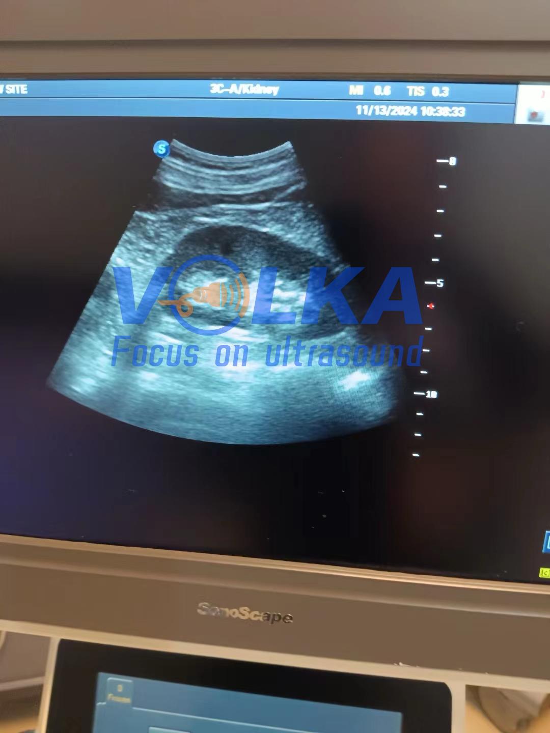
128 elements convex array 3C-A (Abdominal, Obstetrics, Gynecology),
1.0-7.0MHz/ R50mm
192 elements linear array L742 (Vascular, Small parts, MSK etc.),
3.5-16MHz/ 38mm
80 elements phased array 4P-A (Cardiac, Transcranial), 1.0-6MHz
192 elements endovaginal 6V3 (Gynecology, Obstetrics, Urology),
3-15MHz/ R10mm
Style and Performance
Through years of continuous innovation, development, and by giving
priority to our customers’ requirements. Based on the revolutionary
platform, combining SonoScape’s core imaging technologies and sleek
ergonomic design, S40 elevates the image performance to a record
level and satisfies even the most demanding clinical requirements,
significantly expanding the value of ultrasound.
Spatial Compound Imaging
Spatial Compound Imaging utilizes several lines of sight for
optimal contrast resolution, speckle reduction and border
detection, with which S40 is ideal for superficial and abdominal
imaging with better clarity and improved continuity of structures.
Pulse Inversion Harmonic Imaging
The harmonic signals are fully preserved without degradation of the
acoustic information, which makes it possible for S40 to image
high-level details and improve contrast resolution by reducing
noise and clutter in the visualizing of subtle lesions, small
parts, vascular and so on.
μ-Scan
μ-Scan uses real-time image processing algorithm to eliminate
speckle and noise artifacts, enhancing tissue margins and borders
by correcting discontinuity between different regions, allowing
improved visualization of real tissue information.
Auto-Adaptive Imaging Processing
It can automatically adapt the acoustic velocity in different
regions to improve the resolution and contrast.


